

Time-sensitive
Ventricular access may be required on a compressed timeline in acute neurologic emergencies, making a practical bedside workflow essential.
THE SOUNDPASS SOLUTION
Intelligent access. Real-time clarity.
EchoVent is being engineered to acquire ultrasound signals from within the ventricular-access pathway and translate those signals into localization feedback.
CLINICAL CONTEXT
The target can be small, the anatomy can shift, and the clinical moment may leave little room for delay.
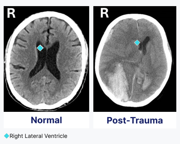
Ventricular access may be required on a compressed timeline in acute neurologic emergencies, making a practical bedside workflow essential.
Conventional bedside placement relies primarily on external anatomical landmarks and does not provide continuous visualization from the advancing catheter tip.
Trauma, edema, and ventricular shift can make the target smaller or move it away from its expected location.
DESIGN OBJECTIVE
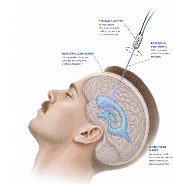
EchoVent brings a miniature ultrasound transducer into the access pathway. The platform is being developed to identify signal transitions associated with anatomical interfaces and translate them into localization feedback while retaining familiar access geometry.
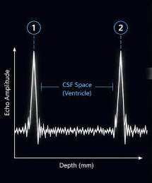
The advancing transducer encounters the near and far ventricular interfaces.
Interface peaks provide a one-dimensional depth signature.
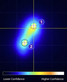
AI-assisted processing allows translation of the one-dimensional signal into a spatial heat map to guide EVD catheter placement in one attempt.
THE ECHOVENT PLATFORM
Each component is being developed as part of a single investigational system for intraprocedural ventricular localization.
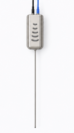
A miniature A-mode ultrasound transducer designed to fit within current EVD catheter luminal diameters and acquire tissue-interface signals during advancement.
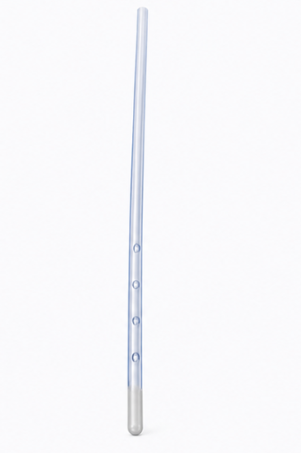
A catheter architecture under development to support acoustic transmission while preserving a familiar ventricular-access workflow.
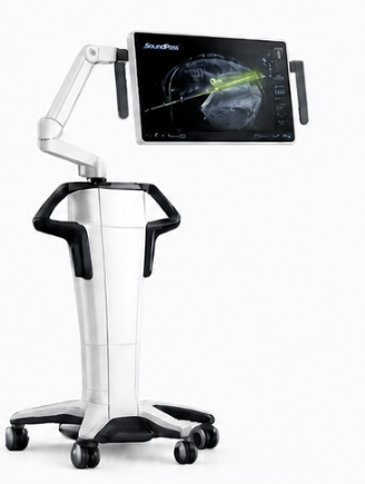
Investigational signal-processing and display software paired with a highly mobile tower designed for positioning over patient beds and surgeon comfort during EVD placement.
DEVELOPMENT PRIORITIES
Designed around the sequence and form factor familiar to ventricular-access clinicians.
Milestone completeA-mode signal acquisition and interpretation have progressed through an initial feasibility milestone.
Milestone completeInitial bench, cadaveric, and animal feasibility work has informed device refinement.
Milestone completeVerification, validation, manufacturing, and regulatory planning remain active workstreams.
PendingCONNECT WITH SOUNDPASS
We welcome inquiries from clinicians, researchers, prospective partners, and investors interested in the SoundPass mission.